


open indirect ophthalmoscope
Re-imaging indirect ophthalmoscopy for the era of open innovation
open indirect ophthalmoscope
Re-imaging indirect ophthalmoscopy for the era of open innovation
Indirect Opthalmoscopy is the most important technique in a ophthalmic clinician's toolbox, allowing them to view desired portions of a patient's retina (the inner tissue of the eyeball which is light sensitive and responsible for vision). This is an invaluable technique to rapidly look for irregularities, pathologies and any other conditions in the retina which may be potentially vision-threatening. These images are termed "fundus" images.


The clinician is able to see a 30 degree field of view at a time. Examination of the retinal tissue can reveal a great deal about the person's present state of health.
Where high-resolution images are required to be taken for diagnosing disorders in the retina, indirect ophthalmoscopy cannot be used. Instead, high-end benchtop devices exist (such as that shown on the right) which capture and record these images. These devices require the patient to be seated and to rest their chin on the device to keep their head in a fixed position relative to the device. A flash of light captures the image after proper alignment is achieved. This is called "fundus photography".

The Problem
The Problem
Most cardiovascular or metabolic disorders show up as vascular pathology on the retina. For instance, Diabetic retinopathy (DR). This is a potentially vision-threatening conditions, where patches of one's retina lose the ability to react to light. This causes patches in one's vision, and if left unchecked, can soon be an irreversible loss of vision. These disorders need to be picked up early and can be treated. However, they require two things - the ability to capture high quality photographs of one's retina, and the ability to interpret those images and diagnose conditions. The first one is done using extremely high quality machines in the market, which are usually very expensive. The second is usually done by a trained clinician (ophthalmologist). Both of these are extremely difficult to get access to in resource-constrained areas or part of large-scale public health screening programs, where they are needed the most.

On the left is a retinal (fundus) image of a normal healthy individual, taken using a high-end benchtop fundus imaging tool. Notice the bright white optic disk on the right, the blood vessels emanating from it (which supply blood to the retina) and the dark spot slightly off center, which is the fovea (responsible for central vision through which we do complex visual tasks like reading).

On the right we have a fundus image of an individual suffering from Diabetic retinopathy. Notice the flaky white spots all over the retina. These "exudates" are deposits which causes "patches" to form in one's vision. Diagnosing this condition is impossible without a good quality fundus imaging camera, which typically costs a few thousand dollars and is extremely large and heavy - definitely not a good solution for field screening programs.

What does this mean for the patient? Those small white spots lead to "blind spots" in one's field of view. The image on the left simulates vision for someone suffering from this condition. If left unchecked, this can lead to permanent vision loss and the inability to perform routine tasks. India, where we are building this product, has the dubious reputation of being the diabetes capital of the world - about 63 million people suffer from diabetes, with this figure likely to go upto 80 million by 2025. About 37% of urban south asians suffer from diabetes or pre-diabetes and are at risk of developing DR. Timely treatment can reduce risks by more than 90%, however symptoms of vision loss only present themselves after significant progression of the disease.
Our Solution
Our Solution
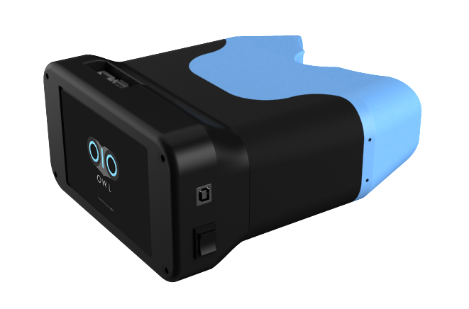
Our product - the Open Indirect Ophthalmoscope - brings all the advantages of the latest technologies into this age-old but elegant technique. The elegant and intuitive form factor brings a new dimension to this technique - portability and new possibilities for image post-processing. Our device enables clinicians to see the same features and level of detail that they are normally accustomed to with the additional portability which enables this powerful technique to be taken out into smaller clinics, field workers and the remotest of locations.
A high-quality PORTABLE IMAGING SOLUTION

A fundus image taken using state of the art clinical instruments

A fundus image taken using our device
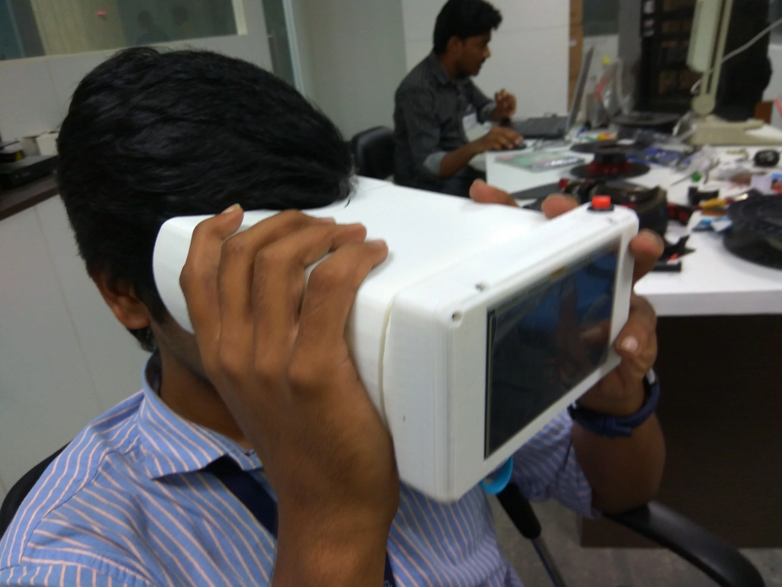
PORTABLE AND ERGONOMIC FORM FACTOR
The device was envisioned as an intuitive handheld device which would require no complex instructions to the patient. Interaction is as straightforward as looking through a pair of binoculars.
integrated machine learning
The device integrates into a massive online grading system (theia) built by graduate students at MIT Media Lab. This system is able to grade the severity of DR in images on a scale of 0 (not present) to 4 (severe). The system uses a deep learning algorithm (convolutional neural networks) and a powerful GPU accelerated machine to be able to process images on cloud in a few seconds. This enables rapid screening and triage without the need for a trained expert to assess each and every image, a powerful tool for eye care clinics and public health programs.

get involved
get involved
build your own
We've put all the instructions to build one in the public domain! Check it out at our Hackaday Page and Instructables Page
order your own
We're still testing the device and it's not in the market yet. However, if you would like to be a beta tester, do get in touch with us!
become a contributor
Are you interested in helping us improve the device? Are you an engineer who's passionate about building open technology with big impact? Send us a mail and follow us on hackaday and github. Send us a mail at srujana [AT] lvpei.org with the subject Open Indirect Ophthalmoscope
THE TEAM
Sandeep Vempati - Project Lead
Dr Jay Chhablani - Principal Investigator
Acknowledgements
Tristan Swedish
Dhruv Joshi
Devesh Jain